We present the case of a 30-year-old Belgian soldier who developed severe leptospirosis after freshwater immersion during a military training exercise in the Vesder River, Belgium. Initially asymptomatic, he presented two weeks later during deployment in Corsica with high-grade fever, intense myalgia, and gastrointestinal symptoms. His condition rapidly progressed to septic shock with hepatic and renal dysfunction, requiring ICU admission. Blood cultures were negative, but Leptospira-specific serology confirmed infection with Leptospira interrogans serovar Semaranga Patoc.
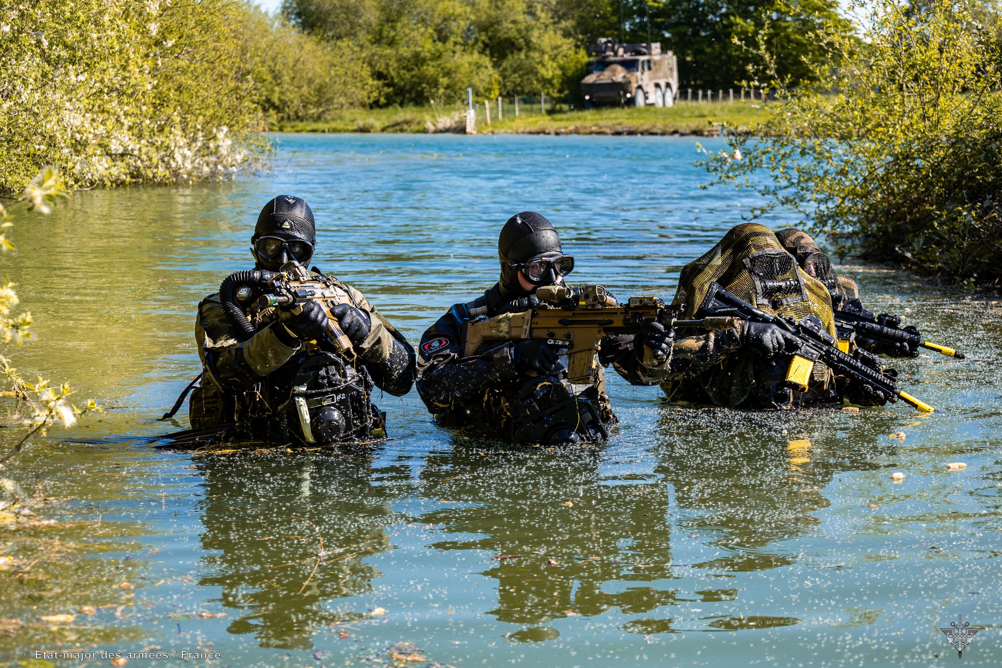
This case highlights the growing relevance of leptospirosis among military personnel exposed to freshwater environments. Although traditionally regarded as a tropical disease, leptospirosis is increasingly recognized in temperate climates, including Belgium. National surveillance data indicate a gradual rise in domestic cases, likely underdiagnosed in non-tropical settings.
Early recognition depends critically on a detailed exposure history. Asking targeted questions about freshwater contact—especially in risk populations such as soldiers—is essential, as prompt empirical antibiotic therapy can be lifesaving when diagnostic tools are unavailable.
Introduction
Leptospirosis is a zoonotic infection caused by pathogenic spirochetes of the genus Leptospira. Humans become infected through contact of skin or mucous membranes with water or soil contaminated by the urine of infected animals, most frequently rodents(1). Although often considered a tropical disease, leptospirosis is now reported worldwide and represents one of the most widespread zoonoses (2).
Clinically, leptospirosis has a broad spectrum. Many cases are mild and self-limiting, but severe disease—classically described as Weil’s disease—can occur, characterized by jaundice, renal failure, pulmonary hemorrhage, and septic shock (3). The case – fatality rate of severe leptospirosis can rise to 52% even with ICU support (4).
The disease is of particular relevance for military personnel. Training often involves activities such as immersion in rivers, swamps, and floodplains, conditions that heighten the risk of exposure (5). Historical outbreaks have been described in U.S., French, and British troops, mostly in tropical or subtropical environments.
In Belgium, the condition is far less frequently reported, yet surveillance data suggest a slow but steady rise in incidence . Climate change will likely further increase the risk of leptospirosis in the future; in a recent report from the European Environment Agency, leptospirosis is listed among the infectious diseases sensitive to climate factors and considered a potential threat to health (EEA) (6).
We describe here the case of a Belgian soldier who developed severe leptospirosis following immersion in the Vesder River. His illness began while deployed in Corsica, and the delayed recognition illustrates both the diagnostic difficulties in austere environments and the importance of exposure history.
The objective of this case presentation is twofold. Firstly, to emphasize the importance for military physicians of obtaining a comprehensive medical history, with specific attention to potential freshwater exposure as a risk factor. Secondly, to highlight the rising incidence of leptospirosis in Belgium.
Case Presentation
A 30-year-old Belgian soldier with no significant medical history participated in a military training exercise involving repeated immersion in the Vesder River near Verviers, an area known for urban runoff and wildlife contamination. The soldier reported ingesting river water during the exercise but remained asymptomatic initially.
Five days later, he developed headache, diffuse myalgia, and malaise, which progressively worsened over the following week. Approximately two weeks after exposure, while deployed in Corsica, he developed a low-grade fever that escalated to 40 °C over several days, accompanied by generalized myalgia—most pronounced in the calves—fatigue, nausea, vomiting, and watery diarrhea. Initially managed as viral gastroenteritis with fluids and paracetamol, his condition deteriorated, and he presented to a civilian hospital with hypotension and tachycardia.
On admission, the patient appeared acutely ill and jaundiced. Vital signs showed a temperature of 38 °C, heart rate 110 bpm, and oxygen saturation of 87% on room air. Physical examination revealed conjunctival suffusion and bilateral crackles on pulmonary auscultation; the abdomen was diffusely tender without hepatomegaly, and mild confusion was noted.
Arterial blood gas analysis showed a pH of 7.42 (normal 7.35–7.45), a pCO₂ of 24 mmHg (normal 35–45 mmHg), and a pO₂ of 72 mmHg (normal 80–100 mmHg), indicating compensated metabolic acidosis with hypoxemia. Laboratory investigations revealed leukocytosis with a white blood cell count of 19.98 × 10⁹/L (normal 4–10 × 10⁹/L), neutrophil predominance, and thrombocytopenia (platelets below 150 × 10⁹/L). C-reactive protein was markedly elevated at >268 mg/L (normal <5 mg/L), and procalcitonin was 99.6 ng/mL (normal <0.5 ng/mL).
Renal function was impaired with a creatinine level of 142 μmol/L (normal 62–106 μmol/L). Hepatic parameters showed a total bilirubin of 30 μmol/L (normal <21 μmol/L) and mild elevation of transaminases. Chest radiography demonstrated bilateral patchy infiltrates suggestive of pulmonary involvement.
Initial working diagnosis was severe community-acquired pneumonia complicated by septic shock. Empirical broad-spectrum antibiotic therapy was initiated with ceftriaxone, metronidazole, and gentamicin, in addition to aggressive fluid resuscitation. The patient was transferred to the intensive care unit (ICU) for advanced monitoring and management.
In the ICU, the differential diagnosis was broadened to include leptospirosis, viral hepatitis, and hantavirus infection, in addition to bacterial sepsis of respiratory or gastrointestinal origin. Antimicrobial therapy was adjusted to cefotaxime combined with doxycycline to provide coverage for atypical pathogens including Leptospira spp. Unfortunately, no data were given regarding neuroimaging or cerebrospinal fluid examination.
Microbiological investigations included blood cultures, serological assays, urinary Legionella antigen testing, and sputum PCR analysis. Importantly, urine PCR was positive for Leptospira DNA, providing strong evidence for leptospiral infection. In consultation with the hospital microbiologist, antimicrobial therapy was subsequently de-escalated to ceftriaxone monotherapy.
Over the following week, the patient demonstrated gradual clinical improvement with resolution of shock and stabilization of renal and hepatic parameters. His pulmonary status improved, and he was successfully weaned off supplemental oxygen. He was discharged from the ICU to a general medical ward for continued supportive care.
Given his military status, arrangements were made for medical repatriation to Belgium, where he was admitted to University Hospital Leuven (UZ Leuven) for ongoing follow-up. At UZ Leuven, serological testing confirmed infection with Leptospira interrogans serovar Semaranga Patoc. This confirmed the diagnosis of severe leptospirosis with multi-organ involvement, including hepatic, renal, and pulmonary manifestations.
Discussion
This case illustrates the potential severity of leptospirosis, even in otherwise healthy young individuals such as military personnel. Leptospirosis is transmitted through direct or indirect contact with the urine or tissues of infected animals. Leptospira spirochetes can enter the body via skin abrasions or mucous membranes, as well as through inhalation of contaminated aerosols. Once inside, the bacteria disseminate hematogenously, leading to leptospiremia and vasculitis, which explain the wide clinical spectrum of the disease (7).
The incubation period averages 10 days (ranging from 5 to 14 days). Clinical manifestations may vary from subclinical infection to a self-limited systemic illness, which accounts for approximately 90 percent of cases, or progress to severe disease (8), the illness evolves in two phases. The initial septicemic phase, lasting 5 to 7 days, is characterized by the presence of Leptospira in blood or cerebrospinal fluid. Symptoms include high fever, headache, myalgias (predominantly in the calves and lumbar region), abdominal pain, nausea, vomiting, diarrhea, cough, and conjunctival suffusion without discharge (9).
The subsequent immune phase may last from 4 to 30 days. During this phase, IgM antibodies are produced and leptospiruria appears. Clinically, patients may present with jaundice, renal failure, respiratory distress, hemoptysis, aseptic meningitis, cardiac arrhythmias, and even pancreatitis. The most characteristic severe manifestation is Weil’s disease, which typically emerges after the acute phase and is marked by hepatic dysfunction combined with renal and pulmonary involvement. Without treatment, the mortality rate of severe leptospirosis can reach up to 52% (10)
A major challenge highlighted by this case is the difficulty of early diagnosis, particularly in resource-limited or austere environments where military forces often operate. The prodromal symptoms of leptospirosis—fever, myalgia (classically in the calves), and headache—are indistinguishable from a wide range of viral and bacterial infections including influenza, dengue, malaria, rickettsial disease, and viral hepatitis (11).
In this patient, the onset of gastrointestinal symptoms led clinicians to suspect viral gastroenteritis, delaying the consideration of leptospirosis until the illness had progressed to a life-threatening state. Such diagnostic delay is not unusual, especially given that clinical features such as fever, thrombocytopenia, and renal impairment are not specific to leptospirosis and are encountered in virtually all forms of septic shock. This underscores the need for heightened awareness in populations at risk, particularly soldiers with freshwater exposure.
Laboratory confirmation of leptospirosis remains problematic in operational settings. Blood cultures are rarely helpful due to the fastidious growth requirements of Leptospira. The gold standard, the microscopic agglutination test (MAT), requires paired sera and specialized laboratory facilities, making it impractical in the field (10). PCR-based methods can offer earlier detection, but their availability in military or rural environments is limited.
Consequently, the diagnosis must often be clinical, driven by epidemiological context and a high index of suspicion. The present case illustrates how a simple but targeted question regarding freshwater immersion could have guided earlier consideration of leptospirosis. Thus, detailed exposure history remains indispensable, particularly in the military population.
For military clinicians, this case emphasizes the importance of maintaining leptospirosis in the differential diagnosis of any febrile soldier with sepsis, renal dysfunction, and thrombocytopenia following field exercises. Such a triad, though non-specific, should trigger specific inquiry into water exposure.
Outbreaks can incapacitate large numbers of troops, and diagnosis in the field is difficult. Misdiagnosis and treatment delays will have potentially fatal consequences. A similar conclusion was reached in outbreaks among U.S. and British soldiers in Panama and Belize, where delayed recognition of leptospirosis contributed to disease severity (12).
For armed forces, leptospirosis poses particular operational challenges. Importantly, military medical training should emphasize that leptospirosis, though historically associated with the tropics, is increasingly relevant in temperate European environments.
Management strategies for leptospirosis are well-established, yet outcomes depend critically on timing. Antibiotics such as intravenous penicillin, ceftriaxone, or cefotaxime are recommended for severe cases, while doxycycline remains the drug of choice for milder disease and as prophylaxis in high-risk deployments. Supportive therapy—including aggressive fluid resuscitation, vasopressor support, and renal replacement therapy—is essential in mitigating mortality. Importantly, mortality in severe leptospirosis can reach 5–15%, and up to 50% in pulmonary hemorrhagic forms, underscoring the value of early recognition and transfer to advanced care facilities (13).
From an epidemiological perspective, leptospirosis is no longer confined to tropical environments. In Belgium, the incidence has been rising steadily over the past two decades, with reports increasingly linked to heavy rainfall, flooding, and recreational water use (6). Climate change, with its associated increase in extreme precipitation events, may amplify this trend. For military forces training in Belgian river systems, such as the Vesder or Ourthe, this represents a tangible occupational health risk. The present case exemplifies how endemic leptospirosis in temperate climates can significantly impact operational readiness, medical resource utilization, and soldier well-being.
In light of these trends, preventive measures are vital in military health policy. Commanders and medical officers should ensure soldiers are informed about the risks of freshwater exposure and employ protective measures when feasible (12). Additionally, strengthening diagnostic capacity through rapid PCR testing in military laboratories could facilitate earlier detection and treatment.
Conclusion
Leptospirosis should be considered in any febrile soldier presenting with hepatic or renal dysfunction following freshwater exposure during field exercises. Early recognition relies on systematic and detailed history-taking, specifically addressing water contact. In austere or prehospital settings, empirical antibiotic treatment based on exposure risk is essential to prevent severe complications and reduce mortality.
References
- Adler B, de la Peña Moctezuma A. Leptospira and leptospirosis. Vet Microbiol. 2010;140(3-4):287-96.
- Costa F, Hagan JE, Calcagno J, Kane M, Torgerson P, Martinez-Silveira MS, et al. Global morbidity and mortality of leptospirosis: a systematic review. PLoS Negl Trop Dis. 2015;9(9):e0003898.
- Levett PN. Leptospirosis. Clin Microbiol Rev. 2001;14(2):296-326.
- Tubiana S, Mikulski M, Becam J, Lacassin F, Lefèvre P, Gourinat AC, et al. Risk factors and prognostic indicators for severe leptospirosis: a retrospective study. PLoS Negl Trop Dis. 2013;7(1):e1991.
- Haake DA, Levett PN. Leptospirosis in humans. In: Adler B, editor. Leptospira and Leptospirosis. Curr Top Microbiol Immunol. Vol. 387. Berlin: Springer; 2015. p. 65-97.
- Sciensano. Epidemiologische surveillance van leptospirose in België. Brussels: Sciensano; 2023.
- Matsui M, Rouleau V, et al. Pathogenesis of leptospirosis. Microbiol Spectr. 2017;5(2).
- Bharti AR, Nally JE, Ricaldi JN, Matthias MA, Diaz MM, Lovett MA, et al. Leptospirosis: a zoonotic disease of global importance. Lancet Infect Dis. 2003;3(12):757-71.
- World Health Organization. Human leptospirosis: guidance for diagnosis, surveillance and control. Geneva: WHO; 2003.
- Picardeau M. Leptospira: the dawn of the molecular genetics era for an emerging pathogen. Nat Rev Microbiol. 2009;7(10):736-47.
- Levett PN. Leptospirosis: a forgotten zoonosis. Lancet Infect Dis. 2001;1(3):151-60.
- Murdoch DR, Woods CW, Zimmerman MD, et al. The etiology of febrile illness in soldiers deployed to tropical regions. Clin Infect Dis. 2004;39(10):1468-74.
- Gouveia EL, Metcalfe J, de Carvalho AL, Aires TS, Croda J, da Silva AQ, et al. Leptospirosis-associated severe pulmonary hemorrhagic syndrome. Lancet. 2008;371(9621):1907-14.
Mit WhatsApp immer auf dem neuesten Stand bleiben!