Report: Nicholas Fernandes ; Nuno Silva
Surgical Approach of a Dentigerous Cyst
Portuguese Armed Forces Hospital – Dental Service
This article relates to a case where through a panoramic radiography a large bone lesion is detected
that had nothing to do with the initial symptoms of the patient. The treatment method and success is
recorded.
Introduction
The dental service of the Armed Forces Hospital, organically represents the top of the pyramid of dentistry in the Portuguese armed forces. It is integrated in NATO ROLE IV facility. It is also responsible for the oral health care of active duty military, their families, and of the Public Security Police (P.S.P) and Republican National Guard (G.N.R). This service is also responsible for preparing the oral health of deployed military in foreign missions, according to NATO standards, and participates in the selection and recruitment of new soldiers. It is also responsible for the oral health of pilots and paratroopers. When the patient visits for the first time our service we do the medical anamnesis and physical examination. After this we request an Orthopantomography which represents an important diagnostic exam, and also an important tool for forensic identification. This article relates to a case where through a panoramic radiography is detected a large bone lesion that had nothing to do with the initial symptoms of the patient.
Reason for Consultation
A 54-year-old woman came to the dental service at the Armed Forces Hospital, and mentioned "I feel discomfort with the prosthesis on the right side" sic. The pain came from the alveolar ridge on the lower premolar area. Through a periapical radiograph we verified the existence of two unerupted premolars.
Clinical History
Healthy patient without smoking or alcohol habits. Patient has denied the use of any kind of medication.
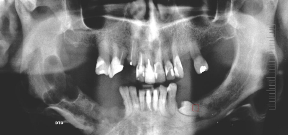
On the first appointment it was requested one Orthopantomography. With this complementary diagnostic exam it was found the inclusion of teeth # 44 and # 45 and a radiolucent lesion involving the # 48 tooth of approximately 3cm, well-defined, unilocular, and compatible with a cystic lesion.
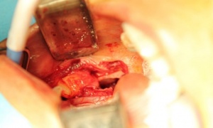
Treatment
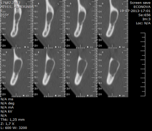
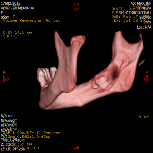
Due to the lesion dimensions, patient was proposed to surgical treatment. Before surgery, it was requested a CT Dental Scan with 3D reconstruction allowing an adequate surgical planning. After consultation of Anesthesiology and collected preoperative conditions, the patient was hospitalized and underwent surgery for third molar extraction, cystic enucleation and extraction of # 44 and # 45. A full thickness flap was performed from the ascending ramus of the jaw to the distal surface of tooth # 43 with discharges. We proceeded to the extraction of tooth and enucleation.
After enucleation of associate injury we have proceeded to irrigation with saline and subsequently repositioned the flap and sutured with silk suture 3/0.
The excised part was sent to the Pathologist for histological analysis. The patient was medicated with Amoxicillin + Clavulanic Acid 875mg + 125mg every 12 hours for 8 days in association with Metronidazol 250 mg every 12 hours for 8 days. Deflazacort was also prescribed 30 mg and Clonixin every 6 hours in SOS. It was recommended a soft diet for a week and the application of ice and 0.12% chlorhexidine gel ont the Post-surgical area. After 8 days the sutures were removed. Follow-up after 1, 3 and 6 months was conducted.
The dentigerous cyst is formed from the accumulation of fluid between reduced enamel epithelium and the crown of an unerupted tooth. It is the second most common type of odontogenic cyst with an occurrence of about 24% compared to all cysts of the jaws and. Is seen more frequently associated with mandibular third molars, maxillary canines and third molar. Are presented mainly as unilateral cysts, unilocular and asymptomatic, episodes of acute pain may occur when there is secondary infection. In tomographic exams, these lesions are presented with well-defined edges and unilocular associated with crowns of impacted teeth, including the cemento-enamel junction. This lesion is usually small however when larger leads to some cortical expansion and a decrease in thickness and can lead to bone fracture. Treatment selection is based on age, size, location, root development, tooth position and relationship with the adjacent teeth and vital structures. Treatment modalities are enucleation or marsupialization.
Diagnosis
Macroscopic lesions and clinical characteristics of the product were consistent with Dentigerous cyst. The enucleated specimen was sent to pathological anatomy for analysis.
According to the anatomopathological report the sample consisted of connective tissue, with rare inflammatory cells, stratified squamous epithelium coated with several layers of cells. Areas with epithelial desquamation were identified.
Anatomopathological diagnosis: dentigerous cyst.
Prognosis
Excellent prognosis when the cyst is enucleated, recurrence is rare.
Conclusion
Complementary diagnostic exams are important both for planning issues either for reasons of identification of potential clinical and pathological situations adjacent to reason for patient consultation. Our patient complained of pain at the level of pre-molars, because the prosthesis exercise trauma inclusion zone. After request of panoramic radiography was found a suggestive radiological image of a dentigerous cyst asymptomatic but with considerable dimensions.
Date: 05/27/2019
Source: Medical Corps International Forum (2/2016)