Report: A. G PIZARRO, E. DE VERA (PHILIPPINES)
Rotary Pilot Low Back Pain in Relation to Occupational Hazards – a Case Report
Rotary AC pilots all over the world have complaint about health problems like low back pain due to factors related to their job.
The objective of the following study is to present factual evidences in support to the claims that chronic low back pain of pilots is caused primarily by vibration and poor posture.
Introduction
It has been postulated that stresses brought about by the nature of the job of rotary pilots cause some health problems, specifically musculoskeletal in nature that are manifested as low back pain (LBP), primarily due to constant vibration of the aircraft, cockpit seat design and posture among others. This case study aims to show the medical history of a typical Philippine Air Force (PAF) rotary aircraft pilot who in his later years has progressively manifested low back pain, which based on theory, may have been induced and further exacerbated by his years of service with the PAF. The significance of the study shall bring forth a clear example of the underlying mechanism of the musculoskeletal problems a typical rotary wing pilot complains of, its possible causes and effects and consequent management and preventive measures one can take so as to minimize if not stop the occurrence of said musculoskeletal problems.
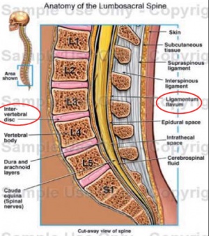 Lumbosacral anatomy, sagittal /
cut away view
C.B. 42-year old, male, Filipino from Pasay City is the subject for discussion in this case. The subject served with the PAF from 1991 finishing his Flying School in 1993, eventually becoming a rotary wing aircraft pilot. Based on records obtained, the subject had 248.5 flying hours in fixed wing aircraft as part of his training as an aviation cadet and later, a total of 1,783.4 flying hours in rotary aircraft giving him an aggregate total of 2,031.9 flying hours as of June 2004, the approximate time when the subject left the service. For his medical history, the subject has no significant co-morbid medical problems during his entire military career, being only confined once at Philippine Air Force General Hospital (PAFGH) in April 2003. He was admitted because of a helicopter crash he was involved in but based on records and interviews with the subject, the subject had no gross or immediate manifestations of any injury other than minor bruises. Familial history of the patient again revealed no significant familial illnesses that may be deemed contributory to a poor physical makeup of the subject. He smokes and drinks occasionally as part of his personal and social history. The subject progressively complained of low back pains since 1999 which were treated by his personal physician symptomatically as possibly musculoskeletal and/or urologic in nature, with the symptoms being relieved by analgesics. The subject left the service in 2004 and shifted to a non flying aviation career overseas wherein he no longer was exposed to the physical stresses of flight. Based on privileged communication with one of the authors, the subject complained of the presence of a dull backache that led to him having a general workup with emphasis on his primary complaints. In January 2010, or almost six years after leaving the service, the subject underwent a series of tests to determine the cause of his progressive low back pains. Magnetic Resonance Imaging (MRI) done on his lower back finally detected the culprit of his illness.
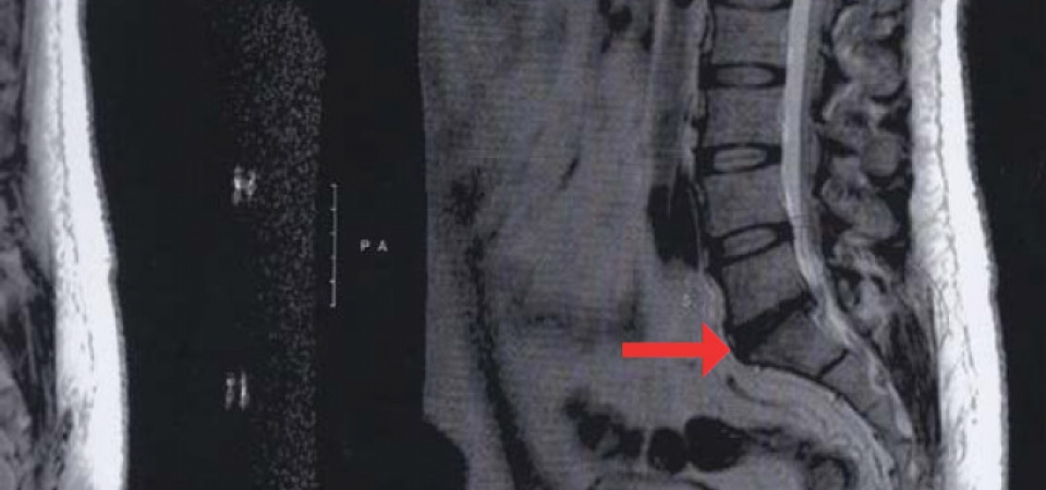
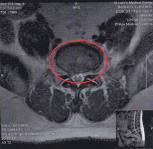
From the above report, there is an anatomical discrepancy with regards to the lumbosacral area of the subject that corresponds to the area of his back pains. Figure 2 shows the actual MRI plate of the subject. Notice the wide gap seen at the space between L5 and S1 intervertebral space as compared to other intervertebral spaces. There is also marked posterior herniation of the L5-S1 intervertebral disc to the spinal cord.
ANALYSIS
From the above mentioned data, the significant inputs one must focus on the significant factors of the subject’s history including the following:
a) history of being exposed to vibratory and postural stresses in a significant number of hours for a relatively short period of time by virtue of his work
b) singular history of direct trauma,
c) early insignificant medical history
d) progressive low back pain complaints, and
e) significant findings on his lumbosacral area.
First a review of the anatomy of the lumbosacral area shows five lumbar bones that join the five fused sacral bones, with muscles and ligaments providing support which are anatomically located at the lower back region just above the buttocks. A sagittal view of said area is shown as reference:
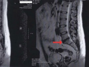 Actual MRI picture of subject
Long hours in the cockpit, ineffective seat padding, poor posture, night vision goggle use, and constant vibration all may contribute to strain and fatigue in the lumbar musculature. Reports also from the U.S Naval Operational Medicine Institute revealed that helicopter pilots reported a higher incidence of LBP as compared to other pilots which was attributed to increased exposure to vibratory forces from rotary wing aircraft. These repetitive vibratory insults to the spinal column specifically to the center of gravity which in the human body is located slightly posterior to the midsagittal plane and at the level of the second sacral vertebrae, are major contributory factors to the cause of indirect trauma to the spinal column and its contents. The fact that our subject has had long hours of sitting in the cockpit as manifested by his being a high timer already predisposes him to be affected by undue stresses at his lumbar musculature. Some studies done to further show the stresses pilots receive that may cause LBP were very specific for the type of aircraft our subject flew in being the UH1H and Bell 205 which are standard PAF rotary wing assets. In an early study, there were significant changes in the way the surface of the back moved during up and down vibration as a result of exposure to the UH-1H, vertically vibrated seated posture, and also significantly, all subjects (male and female) indicated significant increase in pain due to sustained sitting in either static or vibrating UH-1H cockpit conditions. A more detailed explanation on the poor cockpit seat design and its impact on the lumbar musculature can be seen in the following picture:
 2D Echocardiography Report
It can be seen in the above picture that a constantly asymmetrical position does not prevent relaxation of the spinal musculature, thus causing more stress at the lumbosacral region. This leads to spasm of the paraspinous muscles which then eventually gets fatigued and a resultant straightening of the normal body lordotic (curved) postion. Thus, the spine loses its normal curve and the vertebral bodies tend to be closer in front, increasing pressure in the front or anterior part of the intervertebral discs. The nucleus pulposus is then displaced to the rear of the intervertebral space, thereby irritating the nerves it will hit. These actions also flatten and stretch the posterior longitudinal ligament which is very pain sensitive, thereby manifesting the LBP rotary wing pilots like our subject may feel. All helicopter aviators are familiar with the term “helo hunch” which refers to the bent forward posture most helicopter pilots assume while flying. In the low back, this posture converts the normal S-shaped spinal curvature of the spine to more of a C-shape. This shortens the deep spinal muscles and stretches the superficial ones. This is an unstable posture and results in excessive fatigue. In addition, this posture forces the front edges of the vertebrae together, and pulls the posterior edges apart putting uneven pressure on the intervertebral disks. In the neck, the pilot is then forced to additionally hyperextend the neck (nose up) in order to see out of the windscreen. Both of these unnatural positions lead to fatigue, overload, and pain. The structure of the cockpit in itself makes the pilot try to keep his hands on the controls, bending the body to the left due to the position of the collective stick lever, which added to the aforementioned helo hunch, makes the back not rest on the seat thereby adding more strain on the already heavily strained back. The trunk thigh angle therefore becomes about 105 degrees, which is short of the 135 degree angle prescribed to be optimal for a good relaxation of the muscle groups in the lumbosacral area.

Our subject had a history of a crash but based on records available and from interviews with the subject, there were no gross recorded injuries obtained and an initial back pain during the actual crash was felt by the subject as verbalized by him. Nevertheless, the history of a traumatic incident despite absence of gross injuries may have had implications to the LBP the patient felt later on long after his crashing incident. Trauma along with repeated strains or injuries to the spinal area is blamed as a causative factor for disc dessication a term which we can see was diagnosed in the MRI of the patient about seven years after his injury. It has been postulated before that vibration, which is a very strong causative agent for LBP in helicopter pilots, can lead to microtrauma and damage to the intervertebral discs on a molecular level. If the intervertebral discs break down then the back will lose much of its flexibility and may be predisposed to further injury. Injury leads to inflammation, and chronic inflammation can lead to anatomic changes in the shape of the bones themselves. These changes in the anatomy can result in chronic low back pain and added to the history of trauma in our subject, it is quite clear what may have caused the dessicated intervertebral disc.
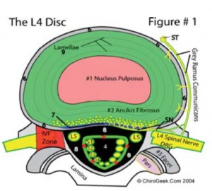 Normal Disc
Going to the medical history of the patient, data revealed insignificant findings during his career in the military and also with his familial history so the issue of prior history of injury or genetic factors can not be clearly established as to causative elements for the LBP that evolved in our subject. His progressive LBP complaints that eventually forced the subject to seek better medical advice than routine out patient consult may have been due to interplay in factors already mentioned. Aging as part of a natural disease process is also highly considered as a cause of the desiccated disc that was seen in his MRI. The most common cause of disc desiccation is still due to the degenerative process. This means that as we age the fluid in the disc evaporates or slowly releases from the disc. This is through normal wear and tear.
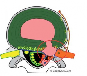
Reading further the impression of the MRI, one can clearly see three distinct causes for the progressive LBP of our subject: disc desiccation which has already been discussed, right posterolateral disc herniation, and laxity of the ligamentum flavum. A disc herniation is the term given to any uneven out-pouching or bulging of the posterior region (back region) of the intervertebral disc as seen on MRI (you can not see disc herniations on X-ray). It occurs when the jell-like center of the intervertebral disc (nucleus pulposus) tears its way through the back-outer portion of the disc (annulus fibrosus) and invades the space (anterior epidural space) where the delicate nerve structures live. And the presence of this nuclear material (which is filled with biochemical irritants called cytokines) in the anterior epidural space may severely irritate these neural structures (i.e., the dura of the thecal sac and nerve roots that make up the giant sciatic nerve), which in turn may cause severe back and/or leg pain.
The nucleus pulposus is intact at the center of the lumbar vertebra and shows no leakage, thus there is structural integrity of the lumbosacral column. Comparing the above normal disc anatomy to an abnormal disc as seen in the figure below,
One can clearly see that the nucleus pulposus has leaked out thus creating pressure at the underlying nerves that causes the pain. Compare this to our subjects MRI, cross-sectional view:
 Typical Herniated Disc
The encirlcled area at the lower left shows a small protrusion that is the nucelus pulposus leaking and putting pressure on the adjacent structures causing the pain felt by the subject. The Ligamentum flavum, the longest ligament in the body,serves as assistance in the maintenance of the erect posture and keeper of the ligament taut during extension. Laxity of the ligaments allows anterior and posterior subluxation of vertebral bodies and pinching of the cord.This could cause the vertebral bones near the weak part of the ligamentum flavum to unduly rub its sharp edges that may further cause pain on movement and consequent limitation or restriction of movement to avoid the pain caused by such actions. So the collective effect of all the aforementioned findings would be pain at the subjects’ low back area with some degree of limitation of movement, all of which were due to the interplay of factors but primarily due to the occupational hazards he experienced during his flying days.
Conclusion
It is evident that the subject, a typical rotary aircraft pilot who served the PAF for about eleven years and have accumulated over 2,000 flying hours eventually complained of severe LBP exemplifies the category of rotary aircraft pilots with the same complaints and subject of studies in other countries. The findings of the MRI done to the subject revealed rather severe damage to his L5-S1 area, which more or less correlates with the human center of gravity that absorbs the vibratory pummeling brought about by the long hours of flight in a rotary aircraft. It is quite safe to assume that the consequent microtrauma and damage incurred by said area had been primarily due to all the exacerbating factors experienced by the subject: prolonged vibration exposure beyond the normal allowable of 2-3 Hertz, poor posturing secondary to aircraft cockpit design constraints, trauma that may have accentuated the degeneration process of his already damaged lumbosacral area, and aging. Though there are other factors like genetics that may also play a role, the fact that our subject is a relatively young individual with no underlying familial history of the disease that may cause the injuries described by the MRI. His history of a singular crash, though traumatic, but with no blatant manifestation of gross injuries nor immediate injures to his lumbosacral area, may also have been responsible for the consequential early weakness of the back musculature that is responsible for providing protection to the lumbosacral area.
 MRI Lumbosacral area, axial view
Having seen the direct effects of the occupational stresses to the health of a rotary aircraft flyer, what comes to mind next are the preventive measures the individual or the organization may take to minimize the deleterious effects of these work induced stressors. Calls have been repeatedly made to suggest improvements in cockpit seat ergonomic designs and the controls to make the workplace of the rotary pilots more “pilot –friendly’ and safer for it could increase visual ability and motor control of the pilots. Vibration control measures that are easily translatable to the user include passive vertical suspension seats that contain a damper and a spring, which can be placed compactly inside the seat cushion or backrest, or non-compactly below the seat cushion or behind the backrest. Simplifying things, a strong pillow or pad that supports the low back area of the pilot can also act as vibration damper. As often mentioned, it is prudent to advise rotary wing pilots to take necessary steps to strengthen the lower back musculature to enhance protection of their low back area, by doing exercises of said area, correct posture at all times even after flights, marinating weight that is proportional to ones’ height to decrease the stress at the affected area and proper body mechanics to avoid undue lumbar stress like when carrying weights or lifting objects. Other easy modalities one can do to prevent the occurrence or worsening of LBP are avoidance of activities that cause undue stress to the low back area, and bed rest to relax the affected musculature. These protective measures must be inculcated early in the minds of the pilots to save their low backs from aggravating injury later in their lives through proper dissemination of information to rotary aircraft pilots by the Air Force Safety Office in coordination with the Office of the Chief Surgeon Air Force and its flight surgeons. For those already with manifesting symptoms of low back pain, in addition to thee aforementioned recommendations, diagnostic exams like MRI be done to localize and determine the extent of injury if any of their lower backs, and prompt treatment with appropriate analgesics and other therapeutic adjuncts to prevent any worsening of the injuries. More new modalities of treatment like platelet enriched plasma therapy are also recommended to be explored.
In conclusion, the existence of low back pain among rotary pilots in the Philippine Air Force has to be studied further, so the aviation community can understand more about the implications and consequences of a simple common complaint of LBP in rotary aircraft pilots. Proper education and preventive measures also have to be set in place institutionally and early diagnosis and treatment be done in order to protect the health and well being of our pilots to maximize their potential and improve their lot. With such, it can be safely said that the advantages of these recommendations will include the following:
- Decreased time lost from work
- Increased combat readiness
- Decreased attrition rates based on chronic pain and injury
- Decreased health care costs
- Overall improvement in the aviator’s health, quality of life and operational effectiveness
Author:
MAJOR JEFFREY ALAN G PIZARRO MC
CAPTAIN ELMA DE VERA MC
Date: 05/13/2019
Source: Medical Corps International Forum (2/2012)


